前瞻技術產學合作計畫
計畫名稱:早期肺癌的相關基因研究
計畫主持人:臺大醫院楊泮池教授
合作企業:Johnson & Johnson Enterprise Innovation, Inc.
成果介紹:
肺癌具有高致死率與發生率,而病例也逐年增加,對原發性非小細胞肺癌患者來說,手術切除術是目前治療最好的方法,然而即使可以接受手術治療,許多病人仍會復發轉移。由於肺癌早期沒有特別症狀,所以對高危險群的長期吸菸者,應定期接受追蹤檢查,例如胸部低劑量輻射電腦斷層掃描,以希望早期發現,才得以在黃金時間接受適當治療。所以如何以非侵入性的方法早期診斷,更近一步或許可以作為全面肺癌篩檢的利器。
在與Janssen的合作案(201912136RIFC)分項A中,我們針對168名病人的早期肺癌組織進行次世代定序。分項B亦針對疑似早期肺癌欲進行手術切除的病人,在手術切除前及術後一週抽取血液病分離其外泌體(exosome),並進行質譜儀分析及次世代定序分析外泌體內所包含之protein及miRNA組成,建立判斷模組以協助早期肺癌之診斷。分項C將針對分項A病人以及公開資料庫的肺癌檢體組織病理數位影像進行專業標註,再運用深度學習方法研究開發早期肺癌組織病理人工智慧輔助診斷系統,並以病人的肺癌生物標記檢測分析結果,建立肺癌組織相關生物標記表現的辨識模型。
技術研發成果
We collected 25 AAH tissue samples, 70 AIS tissue samples, and 73 MIA tissue samples. The pathologist identified the tumor part from the slides (Figure 2). Then, DNA and RNA were extracted from the slides. After library and sequencing by NGS, the data were collected for advanced analysis.

The pathologist identified the tumor part from the slides
Data analysis of the pilot study
First, the nucleic acid amount were all adequate for library. Then, t the expression of gene expression of NGS were adequate for all samples (Figure 3).
Comparing the tumor part with the adjacent normal part, there were 451 up-regulated genes and 1686 down-regulated genes after merging the AIS and MIA tissue samples (Figure 4).

gene expression of NGS

AIS and MIA tissue samples
Then, we explored the relation between tissue samples and mutation numbers. We just select the mutation reads > 5 and allele frequencies > 0.03, and MIA tissue seems have a higher mutation numbers than AIS tissues. The mutation list were listed in Figure 5. The mutation of ERBB2 and EGFR were also detected.

mutation list
The candidate gene mutations associated with the early lung cancer.
We also analyzed the different gene expression associated with the invasion of early lung cancer. The genes, JTB and TMED7, both showed lower expression levels in MIA invasive parts than those in AIS or MIA-AIS parts. CHMP4A showed a higher expression level than the other two types (Figure 6).

different gene expression associated with the invasion
Subgroups in AIS and MIA samples
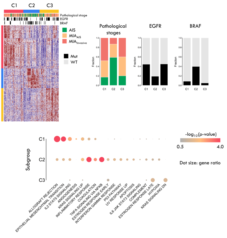
Next, we investigated the transcriptome-based clusters, focusing on AIS and MIA samples. We used non-negative factorization (NMF) to identify the three clusters based on the genes which are highly expressed in AIS or MIA, but lowly expressed in normal tissues. Cluster 1 and 3 (C1 & C3) were majorly MIA samples, but C2 was AIS samples. The function analysis of the cluster-specific genes revealed that immune-related functions were highly expressed in C1 and C2 samples, but C3 did not have significantly enriched functions. We would like to ask whether these subgroups are related to lung cancer progression. Because there were no relapse samples in our cohort, TCGA-LUAD cohort was used to assess the association of subgroups and progression, and we only focused on the stage I-II samples. Based on the cluster-specific genes, TCGA-LUAD were assigned to different subgroups, and we found C3 has better progression-free survival rate than C1 and C2.

Subgroups in AIS and MIA samples
分項計畫C優化第一年計畫所建立的AI輔助標註系統,可依使用情境提升早期肺腫瘤區域至靈敏度達97%以減少漏診/漏標註機會。並藉由AI輔助標註系統協助資深病理專科醫師加速標註工作,取得152片具專業標註的全玻片數位早期肺癌組織病理影像。進一步建立早期肺癌組織病理分類器的演算法程式碼及訓練流程,經校調後的模型分類正常組織/良性腫瘤/侵襲性組織之效能,評估Receiver Operating Characteristic曲線下面積 (AUROC)指標均優於0.98,並可自動量測腫瘤最大直徑供診斷參考(Figure 7)。圖示化驗證模型確實可辨識良性腫瘤/侵襲性組織在病理影像中的位置區域(Figure 8)。(預計) 此外亦建立早期肺癌病理影像生物標記預測模型,至少1項生物標記預測效能AUROC>0.7。
、可視化和腫瘤最大徑量測")
組織病理分類器測試(Patch-level test; N=21)、可視化和腫瘤最大徑量測
、醫師標註(良性腫瘤:綠色;侵襲性組織:藍色)、遮罩與醫師標註之疊圖(黃色/紫色:兩者交集)")
組織病理分類器預測區域與醫師標註區域的圖示化比較,左至右: 熱圖、模型預測之遮罩(紅色)、醫師標註(良性腫瘤:綠色;侵襲性組織:藍色)、遮罩與醫師標註之疊圖(黃色/紫色:兩者交集)
技術特點說明
The retrospectively banked bio-samples of the tumor tissue and adjacent normal tissues will be subjected to a NGS panel examining for whole exomes and RNA sequencing (RNAseq).
The first stage (a pilot study) has been designed to check the feasibility of the whole work flow and to look for potential genetic targets with different mutation pattern or expression level in tumor and normal tissue. A pilot study will be performed first to evaluate the feasibility of normal tissue and tumor tissue for whole exosome sequence and RNA-seq.
Upon success of the pilot study and subject to the decision of Janssen, the second phase of the Project will be initiated which would involve an additional 143 cases.
分項B利用抽血檢驗血液中的外泌體,嘗試建立可預測早期肺癌診斷的生物模組,因此所研發的技術具有方便操作及低侵入性的優點。分項C將開發的早期肺癌組織病理人工智慧輔助診斷系統,期能提供輔助資訊協助並加速組織學診斷,並可從組織病理數位影像上辨識出肺癌相關生物標記的表現,以幫助早期肺癌的診斷與治療決策。
可利用之產業及可開發之產品
本研究成果可用於檢測相關生技產業與智慧醫療產業之商轉及推廣。目前研究仍在進行中,尚無已開發之產品。
推廣及運用的價值:
本技術可合併現有的早期肺癌診斷工具如低劑量電腦斷層,提高診斷的敏感性及特異性,期能提早發現早期肺癌病人並提高其存活率。